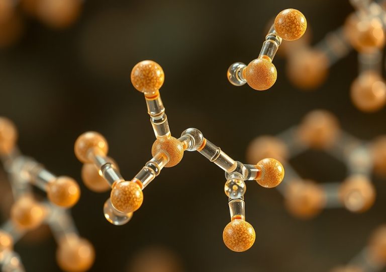

A peptide bond is a covalent amide linkage formed when the alpha-carboxyl group of one amino acid reacts with the alpha-amino group of another, releasing a single molecule of water. This fundamental chemical junction forms the structural backbone of all peptides, polypeptides, and functional proteins. Biochemically, it is not merely a flexible bridge; it is a rigid, resonance-stabilized link that governs how biological macromolecules fold, interact, and function.
While basic chemistry texts define this linkage as a simple single bond, its physical behavior is far more complex. The electronic structure of the bond dictates spatial constraints that restrict how protein chains can maneuver. Understanding these mechanics reveals why Proteins adopt predictable secondary structures like alpha-helices and beta-sheets rather than collapsing into random molecular tangles.
The Molecular Architecture of the Peptide Bond
At its core, the formation of a peptide bond is a nucleophilic acyl substitution classified as a condensation or dehydration reaction. The process begins when the unshared electron pair on the alpha-amino nitrogen ($\text{NH}_2$) of an incoming amino acid attacks the electrophilic alpha-carbonyl carbon ($\text{C=O}$) of a preceding amino acid. This nucleophilic addition forms a tetrahedral intermediate.
Upon collapse of this intermediate, a hydroxyl group is displaced from the carboxyl terminus and picks up a proton from the amino group, departing as a water molecule ($\text{H}_2\text{O}$).
The resulting linkage establishes a characteristic atomic sequence along the polypeptide backbone: $\text{C}_\alpha\text{-C-N-C}_\alpha$. The carbon atom derived from the original carboxyl group retains its double bond to oxygen, forming the carbonyl group, while becoming covalently attached to the nitrogen of the adjacent amino acid. This specific $\text{C-N}$ covalent connection is the amide bond.
Thermodynamically, the synthesis of a peptide bond from free amino acids in aqueous solution is endergonic, possessing a positive change in Gibbs free energy ($\Delta G^\circ’ \approx +8 \text{ to } +16 \text{ kJ/mol}$). Because the reaction is unfavorable under physiological conditions, cells cannot join amino acids spontaneously at meaningful rates.
Biological systems overcome this thermodynamic barrier during translation on the ribosome, where peptidyl transferase—a ribozyme embedded within the 50S or 60S subunit—couples bond formation to the hydrolysis of high-energy nucleoside triphosphates (ATP and GTP) bound to transfer RNAs.
Once formed, the uncatalyzed hydrolysis of a peptide bond in neutral water has an estimated half-life of 350 to 600 years. This extreme kinetic stability ensures that cellular proteins maintain their structural integrity without spontaneously degrading under physiological conditions.
Resonance and Planarity: Why Peptide Bonds Are Rigidity Anchor Points
The defining chemical feature of the peptide bond is its resonance stabilization. The non-bonding lone pair of electrons on the amide nitrogen atom is not localized strictly to the nitrogen nucleus. Instead, these electrons delocalize into the adjacent $\pi$-system of the carbonyl group ($\text{C=O}$).
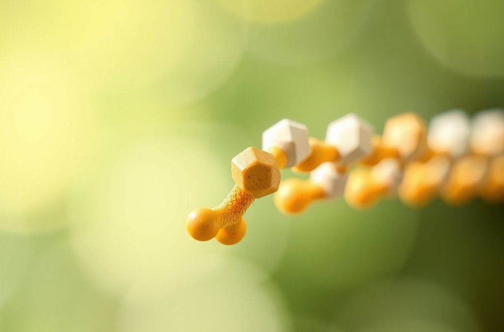
This delocalization can be represented as a resonance hybrid between two principal canonical structures: one with a neutral carbon-oxygen double bond and a single carbon-nitrogen bond, and another with a negatively charged oxygen atom, a positively charged nitrogen atom, and a double bond between the carbon and nitrogen.
Spectroscopic measurements and X-ray crystallography confirm that this delocalization imparts approximately 40% double-bond character to the central $\text{C-N}$ peptide linkage. Consequently, the physical properties of the peptide bond diverge significantly from standard single or double bonds:
| Bond Type | Typical Length (Å) | Rotational Energy Barrier |
|---|---|---|
| Standard $\text{C-N}$ Single Bond | 1.49 Å | Low (~12 kJ/mol) |
| Polypeptide $\text{C-N}$ Peptide Bond | 1.32 Å | High (~80 kJ/mol) |
| Standard $\text{C=N}$ Double Bond | 1.27 Å | Extremely High (>170 kJ/mol) |
Because the bond length of 1.32 Å is substantially shorter than a normal single bond, free rotation around the $\text{C-N}$ axis is severely restricted at body temperature. Overcoming the energy barrier for rotation around this bond requires roughly 80 kJ/mol, which is far greater than the thermal energy available from random molecular collisions. As a direct result, free rotation is virtually eliminated.
This structural restriction forces six continuous atoms to lie within a single, rigid geometric plane: the alpha-carbon and carbonyl group ($\text{O=C}$) of the first amino acid residue, alongside the amide nitrogen, its associated hydrogen ($\text{H-N}$), and the adjacent alpha-carbon of the second residue. This coplanar arrangement, known as the planar peptide group, acts as a structural anchor point in protein chemistry.
Instead of behaving like a flexible chain of independent links, a polypeptide backbone functions as a series of rigid planar tiles connected by flexible hinges.
Trans vs. Cis Isomerism and the Peculiar Case of Proline
Because the double-bond character prevents free rotation around the $\text{C-N}$ bond, the planar peptide group exists in two distinct stereochemical configurations: trans and cis. These isomers are defined by the relative positions of the two neighboring $\text{C}_\alpha$ atoms across the peptide linkage.
In the trans configuration, the adjacent $\text{C}_\alpha$ atoms sit on opposite sides of the $\text{C-N}$ bond (a dihedral angle $\omega \approx 180^\circ$). In the cis configuration, they reside on the same side ($\omega \approx 0^\circ$).
For almost all standard amino acid pairs, the trans isomer is overwhelmingly favored. Thermodynamic studies show that the trans conformation is approximately 8 to 12 kJ/mol lower in free energy than the cis conformation.
This energetic preference stems from steric hindrance: in the cis form, the bulky side chains ($\text{R}$-groups) attached to adjacent $\text{C}_\alpha$ atoms are forced into close physical proximity, creating repulsive van der Waals forces. In non-proline amino acid linkages, this steric strain results in a trans-to-cis ratio exceeding 1000:1.
Proline represents a distinct exception to this rule due to its unique cyclic structure. Unlike the other nineteen standard amino acids, proline’s side chain is covalently bonded back to its own backbone nitrogen, creating a five-membered pyrrolidine ring. Consequently, the nitrogen atom participating in the peptide bond is a secondary amine rather than a primary one.
When any amino acid forms a peptide bond with proline (forming an $\text{X-Pro}$ linkage), the surrounding steric environment changes dramatically. In an $\text{X-Pro}$ bond, whether the conformation is trans or cis, the preceding $\text{C}_\alpha$ atom experiences steric interference from the pyrrolidine ring’s $\text{C}_\delta$ carbon. Because both configurations suffer from comparable levels of steric crowding, the energy difference between trans and cis isomers drops to roughly 2 to 5 kJ/mol.
As a consequence of this minimized energy gap, roughly 10% to 30% of $\text{X-Pro}$ peptide bonds exist in the cis configuration within native protein structures. These cis-proline bonds frequently induce sharp turns, such as reverse $\beta$-turns, which are Essential for compact protein folding.
Because spontaneous uncatalyzed trans-cis isomerization of proline bonds is a slow process that can act as a rate-limiting step in protein folding, cells utilize specialized enzymes called peptidyl-prolyl cis/trans isomerases (PPIases) to accelerate this interconversion.
Torsional Rotations: How Phi and Psi Angles Dictate Protein Folding
While resonance locks the peptide bond itself ($\text{O=C-N-H}$) into a rigid plane, the overall polypeptide backbone retains remarkable conformational flexibility. This mobility occurs because the single bonds connecting the rigid peptide planes to the central alpha-carbons ($\text{C}_\alpha$) are free to rotate. The spatial path of a protein backbone is governed almost entirely by rotations around these two flexible bonds per amino acid residue.
These rotations are defined by two primary backbone dihedral (torsional) angles:
- Phi ($\phi$): The angle of rotation around the single covalent bond between the backbone nitrogen atom and the alpha-carbon atom ($\text{N-C}_\alpha$).
- Psi ($\psi$): The angle of rotation around the single covalent bond between the alpha-carbon atom and the carbonyl carbon atom ($\text{C}_\alpha\text{-C}$).
A third angle, Omega ($\omega$), measures rotation around the peptide bond itself ($\text{C-N}$). However, due to the partial double-bond character discussed earlier, $\omega$ is fixed near $180^\circ$ for trans bonds and $0^\circ$ for cis bonds, rendering it essentially constant.
If all combinations of $\phi$ and $\psi$ angles were stereochemically permissible, a polypeptide chain could adopt an astronomically large number of spatial arrangements. In reality, steric collision limits this flexibility. In 1963, Indian biophysicist G. N. Ramachandran used hard-sphere atomic models to map the sterically allowed combinations of $\phi$ and $\psi$, a visualization now known as the Ramachandran plot.
The Ramachandran plot demonstrates that roughly 75% of all potential $\phi/\psi$ conformational space is forbidden because atomic radii overlap—a state known as steric clash. Unfavorable interactions occur when the carbonyl oxygen, backbone hydrogens, or side-chain atoms approach one another closer than their van der Waals radii permit. The allowed regions are confined to specific clusters:
- Alpha-Helical Region: Clustered in the lower-left quadrant ($\phi \approx -57^\circ$, $\psi \approx -47^\circ$), where right-handed $\alpha$-helices form stabilized networks of intra-chain hydrogen bonds.
- Beta-Sheet Region: Situated in the upper-left quadrant ($\phi \approx -110^\circ \text{ to } -140^\circ$, $\psi \approx +110^\circ \text{ to } +160^\circ$), characteristic of extended strand geometries that align side-by-side into parallel or antiparallel $\beta$-sheets.
- Left-Handed Alpha-Helical Region: Located in the upper-right quadrant ($\phi \approx +57^\circ$, $\psi \approx +47^\circ$), a sparse region populated predominantly by glycine due to its lack of a bulky side chain.
By constraining backbone mobility to these distinct thermodynamic valleys, the physical limits of the peptide bond drive unstructured amino acid sequences to collapse rapidly into stable, predictable secondary structures.
Biochemical Synthesis vs. Hydrolysis: Thermodynamics of Linkage
The condensation reaction that joins two amino acids into a dipeptide is thermodynamically unfavorable under physiological conditions. Forming a peptide bond carries a positive standard Gibbs free energy change (ΔG°’) ranging from approximately +10 to +17 kJ/mol, depending on the specific side chains involved and local ionic strength. Because this process is endergonic, the cell cannot synthesize proteins through spontaneous mass action.
Instead, biological systems couple peptide bond formation to the hydrolysis of high-energy phosphoanhydride bonds.
During ribosomal protein synthesis, this energetic barrier is overcome upstream through aminoacyl-tRNA synthetases. These enzymes utilize ATP hydrolysis to form an aminoacyl-AMP intermediate, which is subsequently transferred to the 3′-hydroxyl group of a transfer RNA (tRNA) molecule. This esterification “charges” the tRNA, effectively storing metabolic energy within an activated aminoacyl ester linkage.
When the ribosome’s peptidyl transferase center—a ribozyme embedded within the 50S (bacterial) or 60S (eukaryotic) large ribosomal subunit—catalyzes the attack of an incoming aminoacyl-tRNA’s α-amino group on the ester carbonyl of the peptidyl-tRNA, the reaction proceeds exergonically without requiring direct ATP consumption at the active site.
Conversely, the reverse reaction—peptide bond hydrolysis—is highly exergonic, with a ΔG°’ of roughly -8 to -12 kJ/mol. Despite this thermodynamic driving force toward degradation, uncatalyzed peptide bond cleavage in neutral aqueous solution at 25°C is extraordinarily slow.
The resonance stabilization of the amide linkage creates a high activation energy barrier (ΔG‡ ≈ 80-100 kJ/mol) against nucleophilic attack by water. Consequently, the uncatalyzed half-life of a typical peptide bond in water is estimated to be between 400 and 1,000 years.
Cells bypass this kinetic stability using proteases. These enzymes employ diverse catalytic strategies—such as the Ser-His-Asp catalytic triad in serine proteases or zinc coordination in metalloproteases—to polarize the carbonyl oxygen, activate a nucleophile, and stabilize the oxyanion tetrahedral intermediate, accelerating hydrolysis rates by factors up to 1016.
Peptide Bond Characteristics vs. Other Biological Linkages
To understand the structural utility of the peptide bond, it must be evaluated alongside the primary covalent linkages that polymerize other biological macromolecules, including nucleic acids, carbohydrates, and lipids.
| Linkage Type | Primary Biomolecules | Geometric Rigidity & Resonance | Thermodynamic Formation Cost (ΔG°’) | Uncatalyzed Half-Life (pH 7, 25°C) |
|---|---|---|---|---|
| Peptide Bond | Proteins, Peptides | Planar; ~40% double-bond character; restricted rotation around C-N bond. | Endergonic (+10 to +17 kJ/mol); driven by ATP-dependent tRNA charging. | ~400 to 1,000 years |
| Phosphodiester Bond | DNA, RNA | Tetrahedral around Phosphorus; flexible backbone rotation around P-O bonds. | Endergonic; driven by cleavage of pyrophosphate (PPi) from dNTPs/NTPs. | DNA: ~30,000 years RNA: ~4 years (due to 2′-OH self-attack) |
| Glycosidic Bond | Polysaccharides, Glycoproteins | Fixed α or β anomeric configuration; flexible chair/boat conformations. | Endergonic; driven by sugar-nucleotide intermediates (e.g., UDP-glucose). | ~5 to 8 million years (for cellulose/starch acetals) |
| Ester Linkage | Triacylglycerols, Phospholipids | Planar ester group, but lower rotational barrier than amide; susceptible to base hydrolysis. | Endergonic; driven by Acyl-CoA thioester high-energy intermediates. | ~1 to 10 years (highly dependent on pH and lipid aggregation) |
Spectroscopic Detection Methods in Modern Biochemistry
Because the peptide bond possesses distinct electronic configurations and vibrational frequencies, biochemists utilize several physical methods to identify, quantify, and characterize polypeptide backbones in structural biology:
- Far-UV Circular Dichroism (CD) Spectroscopy: The peptide bond acts as a chromophore in the far-ultraviolet spectrum (190–240 nm). The n → π electronic transition (around 220 nm) and the π → π transition (around 190–208 nm) are sensitive to the chiral environment created by secondary structures. Distinct CD spectra allow rapid determination of overall α-helix, β-sheet, and random coil content.
- Fourier-Transform Infrared (FTIR) Spectroscopy: The amide backbone exhibits characteristic vibrational modes. The Amide I band (1600–1700 cm-1) arises almost exclusively from the C=O stretching vibration of the peptide bond and serves as a direct readout of secondary structure geometry and hydrogen-bonding strength. The Amide II band (1510–1580 cm-1) reflects out-of-phase C-N stretching and N-H bending.
- The Biuret and BCA Assays: Under alkaline conditions, cupric ions (Cu2+) coordinate with the peptide nitrogens of at least three adjacent amino acid residues, causing a colorimetric shift from light blue to violet (absorbing at 540 nm). In the Bicinchoninic Acid (BCA) assay, the resulting cuprous ions (Cu+) are chelated by BCA to produce an intense purple complex absorbing at 562 nm, providing precise protein quantification based on backbone concentration rather than specific side-chain composition.
Key Takeaways
- Peptide bond synthesis is an endergonic process (+10 to +17 kJ/mol) that relies on coupling to ATP hydrolysis via aminoacyl-tRNA intermediates during ribosomal translation.
- Despite being thermodynamically favorable to hydrolyze, peptide bonds possess an uncatalyzed half-life of 400 to 1,000 years due to the high activation energy maintained by resonance stabilization.
- Compared to phosphodiester and glycosidic linkages, the peptide bond exhibits exceptional planarity and restricted rotational freedom, acting as the structural foundation for secondary protein architecture.
- Biochemists analyze and quantify peptide bond configurations using Far-UV Circular Dichroism, FTIR Amide bands, and copper-coordination assays like the BCA assay.
Watch: Peptide Bond Formation | Animation
Frequently Asked Questions
Why does a peptide bond have partial double-bond character?
A peptide bond has partial double-bond character because the lone pair of electrons on the nitrogen atom delocalizes into the carbonyl system. This resonance hybrid shares electron density between the C=O double bond and the C-N single bond, giving the C-N bond roughly 40% double-bond character. This prevents free rotation around the linkage.
Is peptide bond formation an anabolic or catabolic reaction?
Peptide bond formation is an anabolic reaction. It builds complex macromolecules (proteins) from smaller monomeric units (amino acids). Because it requires a net input of chemical energy delivered through ATP-charged tRNAs to create a covalent bond, it fits the classic metabolic definition of an endergonic anabolic process.
What is the difference between a peptide bond and an amide bond?
A peptide bond is a specific subcategory of an amide bond. All peptide bonds are amide linkages formed between the α-carboxyl group of one amino acid and the α-amino group of another. However, non-peptide amide bonds exist elsewhere in chemistry and biology, such as in synthetic polymers (nylon) or within the side-chain glutamine and asparagine residues.
Why are cis peptide bonds rarely observed in proteins except with proline?
Cis peptide bonds create severe steric clashes between the adjacent Cα carbon atoms and their side chains. In trans configurations, these groups point in opposite directions, minimizing van der Waals strain. Proline is an exception because its cyclic pyrrolidine ring attaches directly to the backbone nitrogen, creating comparable steric hindrance in both cis and trans forms.
How do proteases break peptide bonds if they are kinetically stable?
Proteases break peptide bonds by deploying nucleophilic groups (like activated serine hydroxyls, cysteine thiols, or water molecules coordinated by metal ions) to attack the carbonyl carbon. This bypasses the high activation energy barrier by destabilizing the resonance hybrid and forming a short-lived tetrahedral intermediate, accelerating the hydrolysis rate millions of times over.
Conclusion: The Master Linkage of Structural Biology
The peptide bond is the primary structural linkage of biological systems, combining chemical stability with localized spatial rigidity. By balancing a high kinetic barrier to spontaneous hydrolysis with predictable planar geometry, this covalent amide bond allows linear sequences of amino acids to reliably fold into functional three-dimensional proteins.
For students and Researchers seeking to deepen their understanding, reviewing Ramachandran plots and studying enzyme active-site kinetics offer the most direct path to mastering backbone dynamics. This content is provided strictly for educational purposes, and any applied research or clinical interpretations should be evaluated alongside a qualified biochemist or clinical specialist.