

The core peptides vs proteins difference lies in their molecular size, structural complexity, and conformational stability. Both are polymers constructed from amino acids linked by covalent peptide bonds, but molecules containing fewer than 50 amino acids are classified as peptides, whereas chains exceeding 50 amino acids are designated as proteins.
This arbitrary boundary marks a profound shift in physical behavior: peptides generally function as flexible, unstructured signaling molecules, while proteins fold into rigid, highly specific three-dimensional architectures capable of performing complex mechanical and enzymatic work.
While basic biological definitions focus entirely on chain length, this distinction overlooks the profound biochemical and pharmacokinetic consequences of molecular scale. Understanding how molecular weight influences tertiary structure, enzymatic degradation, cell membrane permeability, and receptor activation is essential for evaluating peptide therapeutics, protein biologics, and dietary supplements. This review examines the exact biophysical boundaries that separate these two classes of biomolecules.
The Molecular Divide: Amino Acid Chain Length and the 50-Residue Boundary
At the most fundamental chemical level, peptides and proteins are constructed from the same building blocks: L-alpha-amino acids joined through condensation reactions that form amide bonds, historically termed peptide bonds. During this reaction, the carboxyl group of one amino acid reacts with the alpha-amino group of another, releasing a molecule of water.
The resulting carbon-nitrogen link possesses partial double-bond character due to resonance stabilization, restricting rotation around the peptide backbone and establishing a planar geometry for each link in the chain.
The scientific consensus established by the International Union of Pure and Applied Chemistry (IUPAC) sets the dividing line between peptides and proteins at approximately 50 amino acid residues. Below this threshold, molecules are categorized based on their specific chain length:
- Dipeptides and Tripeptides: Chains of two or three amino acids (e.g., carnosine, glutathione). These small molecules often utilize specialized active transport systems, such as the PEPT1 transporter in the human intestine.
- Oligopeptides: Chains typically comprising between 4 and 20 amino acids. Examples include peptide hormones like oxytocin (9 residues) and vasopressin (9 residues).
- Polypeptides: Unbranched chains containing between 20 and 50 amino acids. While longer than oligopeptides, they generally lack a fixed, stable tertiary structure in physiological solution.
Molecular weight directly mirrors this chain length classification. Peptides typically exhibit a molecular mass below 5,000 Daltons (5 kDa). For context, insulin—comprising 51 amino acids across two linked chains with a total mass of roughly 5.8 kDa—occupies the historical boundary region between a complex polypeptide and a small protein. Proteins, by contrast, range from approximately 6 kDa to massive macromolecular complexes like titin, which spans over 33,000 amino acid residues and exceeds 3.8 million Daltons.
This mass differential is not merely academic. Molecular weight directly dictates hydrodynamic radius, diffusion rates in extraCellular matrices, and the mechanism by which the human body metabolizes and clears the molecule from circulation.
Conformational Flexibility vs. Higher-Order Folding: How Structure Dictates Function
The defining functional divergence between a short peptide and a fully realized protein is the presence of stable, autonomous tertiary structure. The spatial arrangement of these molecules is categorized into four hierarchical levels: primary (amino acid sequence), secondary (local spatial arrangements such as alpha-helices and beta-sheets), tertiary (overall three-dimensional architecture of a single polypeptide), and quaternary (assembly of multiple polypeptide subunits).
Short peptides lack the thermodynamic driving force required to maintain a permanent three-dimensional shape. Because a chain of 10 to 30 amino acids lacks a sufficiently large hydrophobic interior, it cannot offset the conformational entropy lost during folding. Consequently, in an aqueous environment, peptides exist primarily as dynamic ensembles of rapidly interconverting configurations, often referred to as random coils.
When a peptide binds to its target receptor, it undergoes induced-fit conformational selection, adopting a structured state only upon physical engagement with the binding pocket.
Proteins achieve thermodynamic stability by driving hydrophobic amino acid side chains (such as leucine, isoleucine, valine, and phenylalanine) away from water molecules into a tightly packed internal core. This hydrophobic collapse, combined with intramolecular hydrogen bonding, electrostatic salt bridges, and Covalent disulfide bonds between cysteine residues, locks the protein into a precise tertiary structure. This rigid 3D conformation creates highly specialized functional sites, such as:
- Enzymatic Active Sites: Catalytic pockets configured to lower the activation energy of specific chemical reactions (e.g., polymerases, proteases).
- Allosteric Regulatory Domains: Distant binding sites that alter the protein’s overall shape and activity when engaged by effector molecules.
- Structural Scaffolds: Dense, fibrous networks like collagen and keratin that provide mechanical integrity to tissues.
If a protein loses this specific 3D architecture through thermal or chemical stress—a process known as denaturation—it generally loses its biological function entirely. Peptides, lacking a fixed tertiary fold to begin with, are largely immune to classical structural denaturation, though they remain susceptible to chemical degradation of their individual amino acids.
Watch: Peptides vs Proteins: What's the Real Difference? (Animated)
Bioavailability, Stability, and Half-Life: Pharmacokinetics of Peptides vs. Proteins
The structural disparities between peptides and proteins dramatically dictate their pharmacological behavior, metabolic stability, and administration routes within the human body. The table below outlines the core pharmacokinetic parameters separating small peptides from large globular proteins.
| Pharmacokinetic Parameter | Peptides (< 50 Amino Acids) | Proteins (> 50 Amino Acids) |
|---|---|---|
| Primary Enzymatic Cleavage | Rapid degradation by endopeptidases and amino/carboxypeptidases in plasma and mucosal tissues. | Slower initial cleavage; often internalized via endocytosis prior to lysosomal degradation. |
| Elimination Route | Rapid renal filtration through the glomerulus (cutoff threshold approx. 30-50 kDa). | Hepatic clearance, reticuloendothelial system (RES) uptake, and receptor-mediated endocytosis. |
| Circulatory Half-Life | Very short (minutes to hours) in native un-modified forms. | Extended (hours to weeks; e.g., IgG antibodies persist up to 21 days). |
| Cell Membrane Permeability | Low, but short sequences (e.g., cell-penetrating peptides) can cross membranes via active endocytosis or direct translocation. | Negligible direct membrane permeability; relies entirely on receptor-mediated transport or endocytosis. |
| Thermal & Physical Stability | High resistance to physical shear stress and heat-induced conformational denaturation. | Highly sensitive to aggregation, temperature changes, freeze-thaw cycles, and mechanical shear. |
| Common Stabilization Strategies | N-terminal acetylation, C-terminal amidation, D-amino acid substitution, cyclization, lipid conjugation. | PEGylation, Fc-fusion, glycosylation engineering, human serum albumin binding. |
Because native peptides lack globular shielding, their peptide bonds are exposed to ubiquitously expressed brush-border and plasma peptidases. Consequently, unmodified peptides like native GLP-1 or somatostatin exhibit plasma half-lives of less than three minutes. To overcome this limitation, pharmaceutical chemists introduce modifications such as backbone cyclization (which hides terminal groups from exonucleases) or fatty acid lipidation (which promotes reversible binding to human serum albumin, delaying renal filtration).
Proteins benefit from structural mass that prevents passive renal filtration through the kidney’s glomerular basement membrane. However, their physical fragility presents unique formulation challenges. Exposure to agitation, hydrophobic surfaces, or elevated temperatures can cause proteins to unfold and form irreversible, non-functional aggregates. Additionally, because of their large surface area and foreign epitopes, protein therapeutics carry a significantly higher risk of triggering neutralizing anti-drug antibody responses compared to short, synthetic peptides.
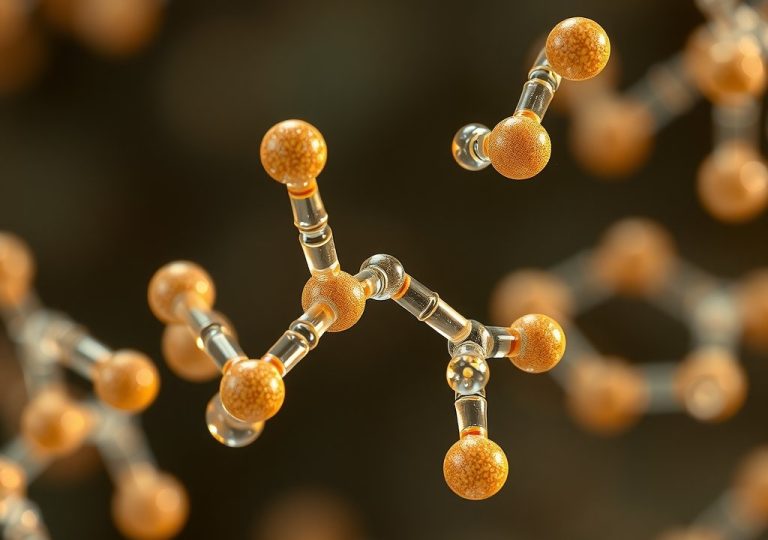
[…] comparing peptide vs protein size, the traditional boundary is set at 50 amino acids: chains with fewer than 50 amino acids are […]